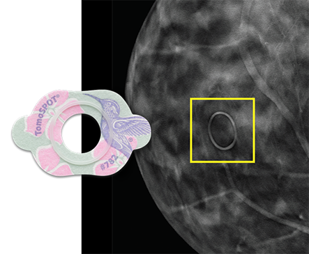
TomoSPOT® for Digital Breast Tomosynthesis
TomoSPOT skin markers are tested and proven to communicate with minimal distraction in digital breast tomosynthesis, or 3D mammography. Digital Breast Tomosynthesis reveals greater tissue detail in high resolution slices, increasing the number of images a radiologist must review.
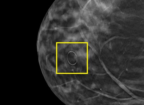
Mole Markers Help Eliminate Questions; Reduce Avoidable Callbacks
Moles, also referred to as skin lesions, often present an interpretation challenge in breast imaging due to the inconsistency with which they image. A mole may image on one view and not the next or it may be absent on the prior year, but image on the current mammogram, and this causes questions as moles can sometimes be confused with intraparenchymal masses. It's not unusual for patients undergoing screening mammography to be called back for additional imaging due to an unmarked mole. The American College of Radiology (ACR) recommends that breast imaging facilities "should require consistent use of radiographically distinct markers to indicate palpable areas of concern, skin lesions, and surgical scars."

Nipple Markers
Why Mark Nipples in Mammography?
How nipple markers help with image quality and image interpretation
As the primary visible landmark on most breasts, the nipple takes on an important role when it comes to screening and diagnostic mammography. Knowing exact nipple location helps eliminate guesswork when it comes to image assessment and interpretation and, more importantly, helps triangulate the location of abnormalities for additional imaging and further workups. However, the nipple isn't always identifiable. Every patient's breasts are unique - some women have inverted nipples, some women have nipples that try as you may, will not go into profile. It may be rolled under the breast, it may point to the side. Nipple location on the image may vary from year to year based on the positioning technique of the individual mammography technologist.
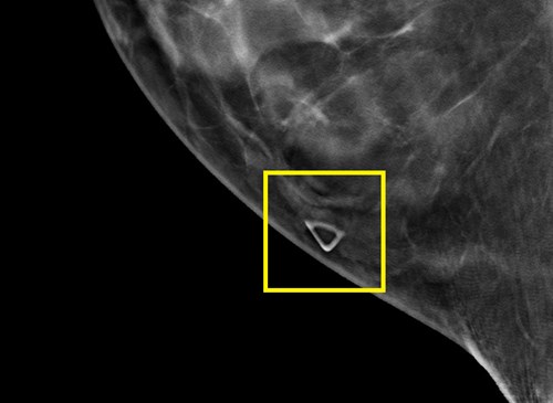
Palpable Mass Markers
Warning! Palpable Lump Felt Here
Radiographically distinct; unmistakable meaning
In 2018, the American College of Radiology (ACR) amended their Practice Parameters for the Performance of Screening and Diagnostic Mammography to recommend that "Facilities should require consistent use of radiographically distinct markers to indicate palpable areas of concern, skin lesions, and surgical scars."
This recommendation aligns perfectly with the mission behind the MED Tech AS
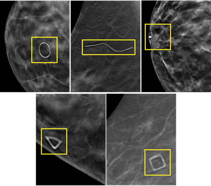
Skin Marking System® for Mammography - 5 distinct shapes to visually communicate and document specific areas of interest or concern to the radiologist directly on the patient's images.
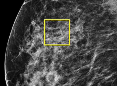
Communicating Focal Pain in Mammography
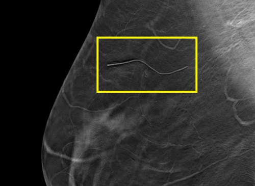
The square marker facilitates correlation between patient-reported symptoms and imaging findings Best practices in mammographic skin marking require consistent use of each distinctly shaped marker to communicate one specific area of concern: the circle to communicate raised moles, the line to communicate scars from previous surgeries, the pellet for indicating nipple location, and the triangle to indicate a palpable lump. So what do you do when a patient complains of focal pain, or has another non-palpable topographic finding that may image, such as a bug bite or cyst? Reusing any of the shapes above could cause confusion and result in inaccurate interpretation and unnecessary additional imaging.
Eliminate Confusion Caused by Architectural Distortion in Mammography
Scar markers act as a reference point to identify the location of previous surgeries With the advent of digital breast tomosynthesis (DBT), radiologists are seeing more architectural distortion from older benign surgeries than they had previously in FFDM, necessitating the need to look back at years of previous year images to determine if the architectural distortion on the DBT image is correlated to a previous surgery or is something new.
A Simple, Effective Way to Improve Efficacy, Reduce Callbacks
Skin markers offer a simple, effective way to improve efficacy and reduce call backs in mammography
No matter what imaging equipment your breast center uses, image interpretation can be faster and more accurate with the consistent use of skin markers. Five distinct shapes subtly, yet clearly, communicate and permanently document the location of moles, skin scars from prior surgeries, palpable masses, nipple position, focal pain or other non-
- Every patient deserves the most breast tissue possible on their image. Bella Blankets protective coverlets provide mammography technologists an extra helping hand by:
- Duis aute irure dolor in reprehenderit in voluptate velit.
- Ullamco laboris nisi ut aliquip ex ea commodo consequat. Duis aute irure dolor in reprehenderit in voluptate trideta storacalaperda mastiro dolore eu fugiat nulla pariatur.
Ullamco laboris nisi ut aliquip ex ea commodo consequat. Duis aute irure dolor in reprehenderit in voluptate velit esse cillum dolore eu fugiat nulla pariatur. Excepteur sint occaecat cupidatat non proident, sunt in culpa qui officia deserunt mollit anim id est laborum

A proactive routine skin marking protocol in mammography
- Aligns with current ACR recommendations
- Reduces risk of false negatives and false positives.
- Reduces unnecessary additional views and callbacks
- Provides visual documentation of breast anatomy from year to year and when transferring images
The MED Tech AS Skin Marking System for Mammography - Booklet
DownloadBreast Cancer Awareness Poster - Every Breast is Unique, Like a Fingerprint
DownloadThe 5 Shape Skin Marking System for Mammography - Mini Poster
DownloadTomosynthesis and Skin Markers, Why Marking Remains a Best Practice in Advanced Breast Imaging
Download